Sign in
 Create Account


Clinical Context in Podiatric Practice
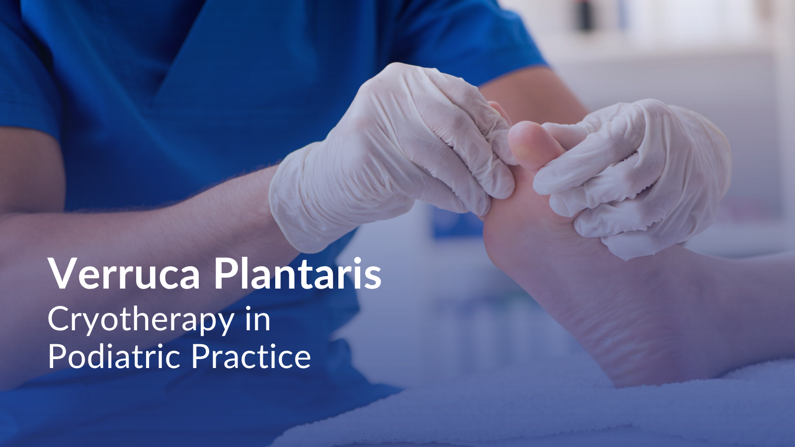
Verruca plantaris is a cutaneous lesion caused by human papillomavirus (HPV) infection and frequently encountered in podiatric practice. Lesions typically develop on weight-bearing plantar surfaces and may become symptomatic due to mechanical pressure and local inflammation.
Clinically, verrucae often present as hyperkeratotic lesions with interruption of dermatoglyphic lines and punctate capillary thrombosis. Pain may be elicited with direct pressure or lateral compression, particularly when the lesion extends deeper into the plantar dermis.
Initial management strategies may include topical keratolytic agents, chemical destruction, sharp debridement, or other ablative approaches depending on lesion characteristics and practitioner assessment.
Anatomical and Tissue Considerations
Unlike neural pathologies, verruca plantaris originates within the epidermal layer, though chronic lesions may extend into the superficial dermis.
In weight-bearing regions, overlying callus formation may obscure lesion margins and increase apparent depth. Plantar skin thickness and underlying fibrofatty tissue density influence both procedural access and cryogenic penetration.
Although the lesion itself is cutaneous, plantar sensory innervation contributes to pain perception, particularly in areas of repetitive loading.
Careful evaluation of lesion boundaries, depth, and surrounding tissue integrity is central to procedural planning.
Procedural Considerations in Cryotherapy
Cryogenic management of verruca plantaris involves controlled freezing of pathologic tissue within defined anatomical limits.
The procedural objective is localized cryonecrosis of infected epidermal tissue while minimizing collateral damage to surrounding healthy structures.
Application is directed toward the lesion surface or through targeted cryogenic delivery depending on lesion thickness and anatomical context. Attention is given to:
- Accurate identification of lesion margins
- Consideration of plantar skin thickness
- Awareness of depth relative to underlying soft tissue
- Controlled limitation of lateral tissue spread
Cryogenic exposure induces intracellular ice formation and vascular stasis within the targeted area, contributing to tissue destruction. The extent of tissue effect depends on depth and duration of freezing, both of which are determined by practitioner judgment.
Post-application tissue response may include localized edema, blister formation, or desiccation consistent with cryogenic tissue injury. Healing progression varies depending on lesion size and anatomical location.
Educational Workshop Excerpt: Cryogenic Application in Verruca Plantaris
This short excerpt was recorded during a podiatric training workshop with Dr Brian Allen, podiatrist, and provides visual context for the procedural considerations discussed above.
The full procedural demonstration are available through the Professional Educational Series for Podiatrists. Request Access
Verruca plantaris represents a localized viral pathology frequently managed in podiatric practice. Cryogenic application constitutes one interventional approach aimed at controlled tissue destruction within defined anatomical boundaries.
Professional Educational Series for Podiatrists
This four-week educational video series, delivered by email, presents cryoanalgesia procedures relevant to office-based podiatric practice.
The program includes clinical discussions on:
- Plantar fascia–related pain
- Morton’s Neuroma
- Foot and ankle bursitis
- Verruca
The educational series is intended exclusively for licensed healthcare professionals.
Request Professional Access
Procedural environment
Procedures illustrated on this page were documented during a podiatric training workshop using the Cryo PainBlocker™ cryoanalgesia system.
This article is provided for healthcare professionals only and for educational purposes.
It does not constitute clinical recommendations, treatment protocols, or outcome claims.
Explore related articles
– Cryoanalgesia for Plantar fascia
– Cryoanalgesia for Foot and Ankle Bursitis
– Cryoanalgesia for Morton’s Neuroma