Sign in
 Create Account

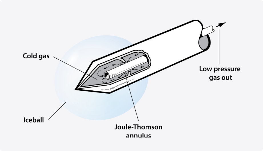
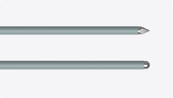
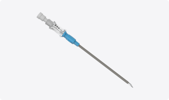
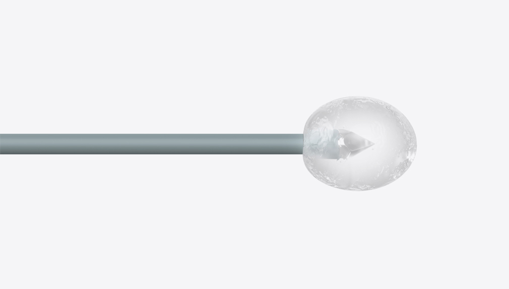
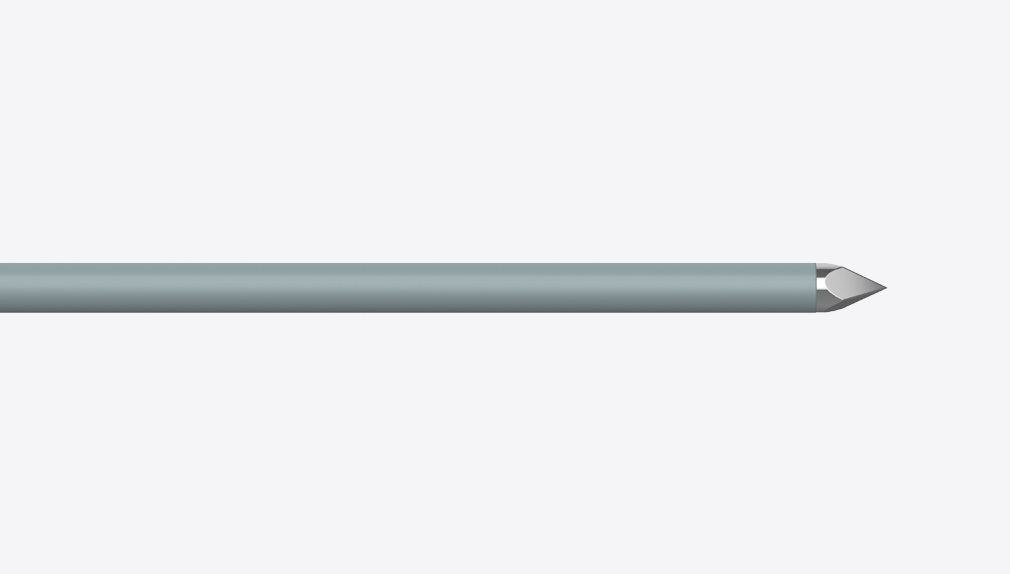
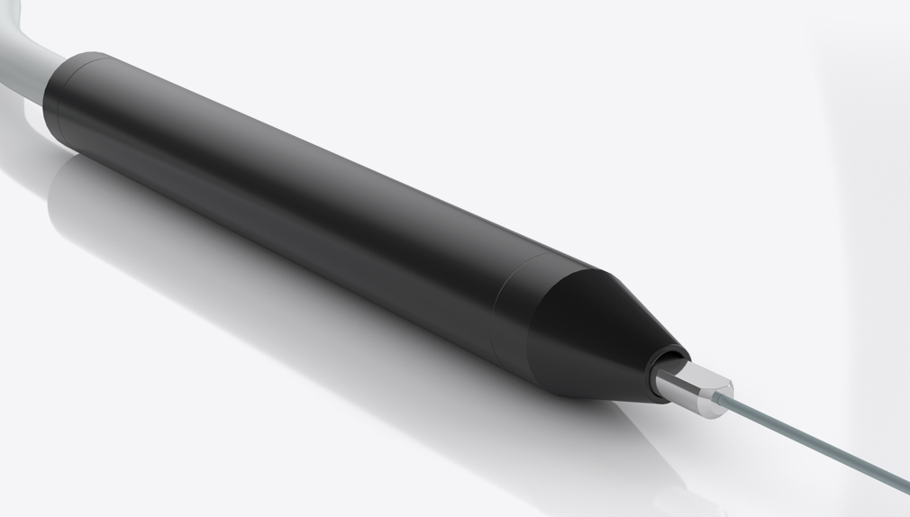
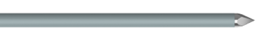
Trocar Probe
The sharp, pyramid-shaped tip of the trocar probe allows for easy access.
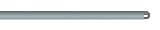
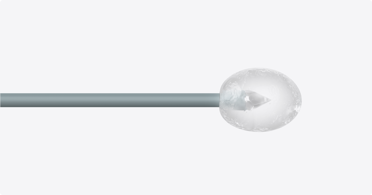
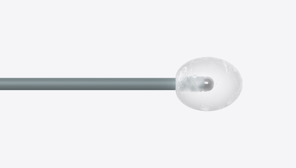
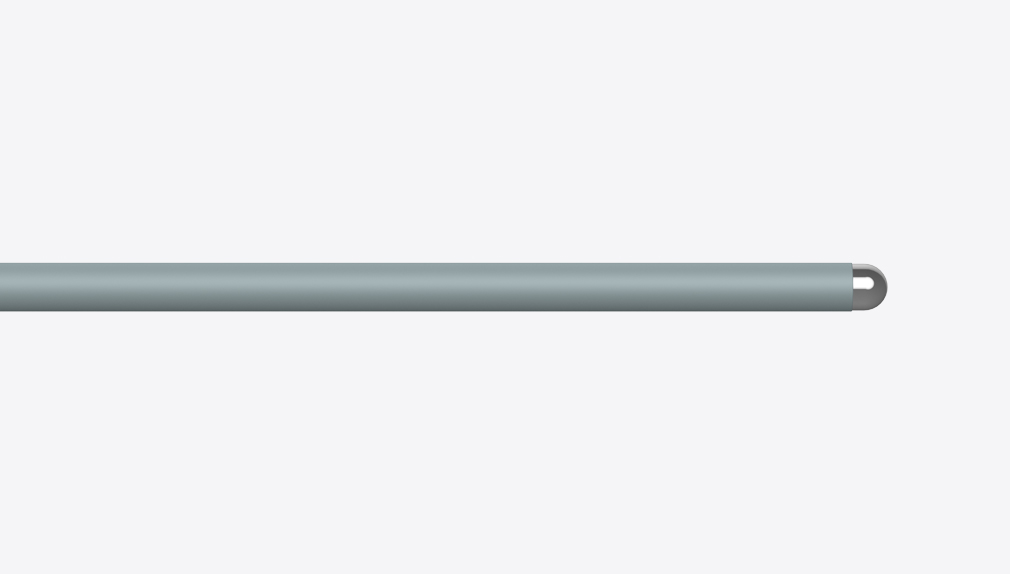
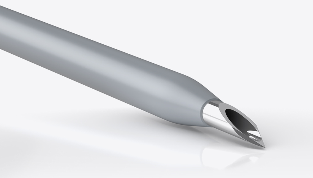
Hemispherical Probe
The blunt tip of the hemispherical probe is designed to minimize nerve and tissue trauma.
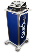
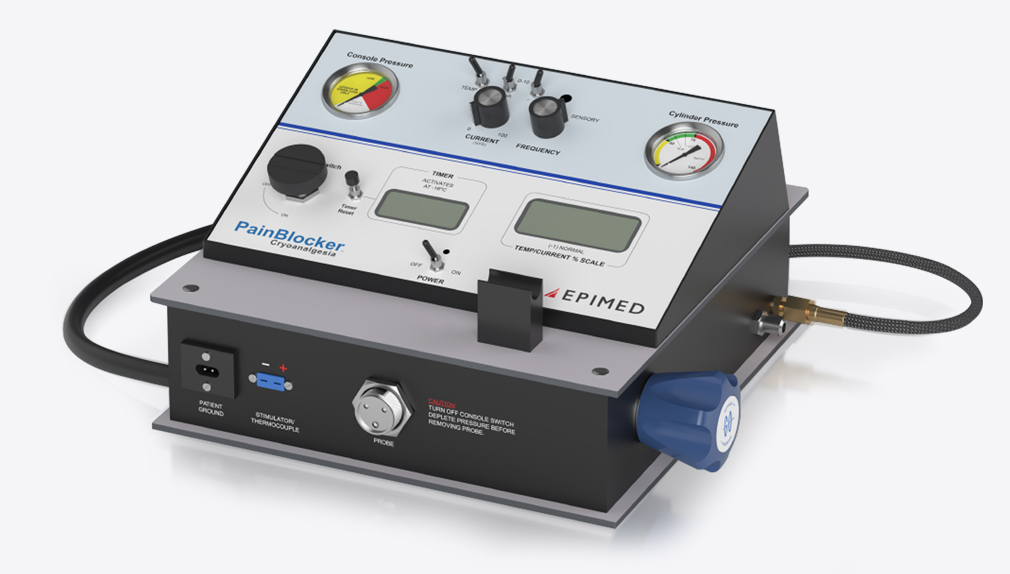
PainBlocker™ Mobile Cart
The PainBlocker™ mobile cart is equipped to hold the PainBlocker™ console, a CRYO probe, and a 20 lb N2O or CO2 cylinder